The physiological root of this phenomenon sits inside the back wall of the eye. Retinal tissue contains roughly 6 million to 7 million human eye cone photoreceptors split into three distinct categories: Short (S-cones, tuned to blue around 420 nanometers), Medium (M-cones, tuned to green around 530 nanometers), and Long (L-cones, tuned to red around 560 nanometers).
These photoreceptors do not operate as razor-sharp digital tuners. Their absorption curves overlap broadly across the visual spectrum. When light from a true single-wavelength source hits the retina, such as a spectral yellow beam at 580 nanometers, it lands right between the peak sensitivities of the M-cones and L-cones. That monochromatic beam stimulates both cone groups at once, prompting the optic nerve to send dual electrical bursts upward.
Now consider what happens when a digital monitor emits a pair of separate wavelengths: one red and one green. The red photons stimulate the L-cones. The green photons stimulate the M-cones. To the visual cortex, the incoming neural code is virtually indistinguishable from true spectral yellow. The eye lacks the mechanical architecture to dissect the incoming wave packet into individual frequencies. It sees dual stimulation and reports "yellow."
| Visual Domain | Physical Medium | Dominant Mechanism | Visual Outcome |
|---|---|---|---|
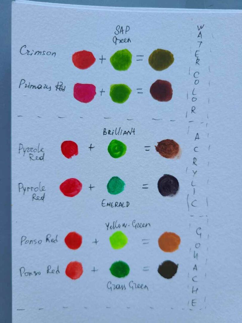
| Pigment & Paint | Oils, acrylics, inks, printing dye | Subtractive wavelength absorption | Desaturated, muddy brown or dark olive |
| Direct Optics | LEDs, OLED displays, theatrical spot beams | Additive photonic accumulation | Radiant, highly saturated yellow |
| Monochromatic Wave | Prism refraction, laser emission (580 nm) | Single-frequency cone excitation | Pure spectral yellow |
| Four-Color Process | CMYK commercial half-tone print | Micro-dot subtractive layering | Near-black chromatic neutral |